Order of Human Back Muscles From Superficial to Deep
28
Back Muscles
The back is the posterior part of the trunk extending from the neck to the pelvis and includes the skin, fascia, muscles, vertebral column, spinal cord, and supporting neurovasculature.
Bones
Bony support for the back comes from the vertebral column and posterior rib cage. The vertebral column extends from the base of the skull down to the tip of the coccyx (see Chapter 26). It protects the spinal cord, supports the weight of the trunk, and transmits this weight to the pelvis and lower limbs. The back is also supported posteriorly by many strong muscles that attach to the vertebral column. The vertebrae, their processes, and the ribs are attachment sites for the back muscles.
The thoracolumbar fascia, like the nuchal fascia in the neck region, is a thick downward extension of fascia that is attached to each of the vertebral spines inferior to TVII, the supraspinous ligaments, the medial crest of the sacrum, and the ribs. It splits into two layers (anterior and posterior layers) and supports and provides attachment for the deep back muscles.
Muscles
The muscles of the back (Table 28.1) are divided into three groups:
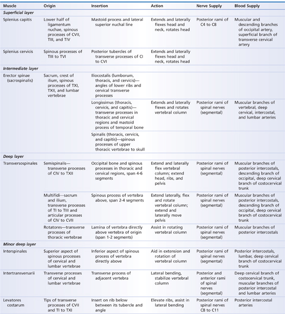
TABLE 28.1 Deep Intrinsic Back Muscles
- The superficial extrinsic back muscles (trapezius, latissimus dorsi, levator scapulae, and rhomboids, Fig. 28.1) connect the upper limbs to the trunk and control limb movements (see Chapter 17).
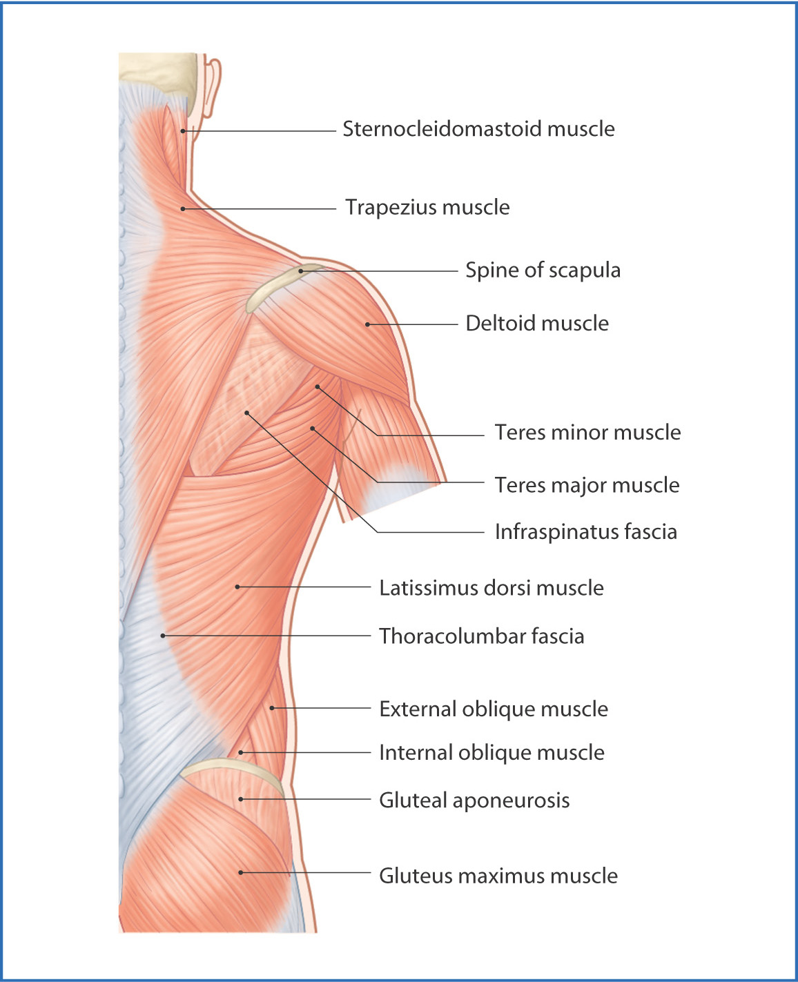
FIGURE 28.1 Superficial muscles of the back.
- The intermediate extrinsic back muscles (serratus posterior, Fig. 28.2) are superficial respiratory muscles.
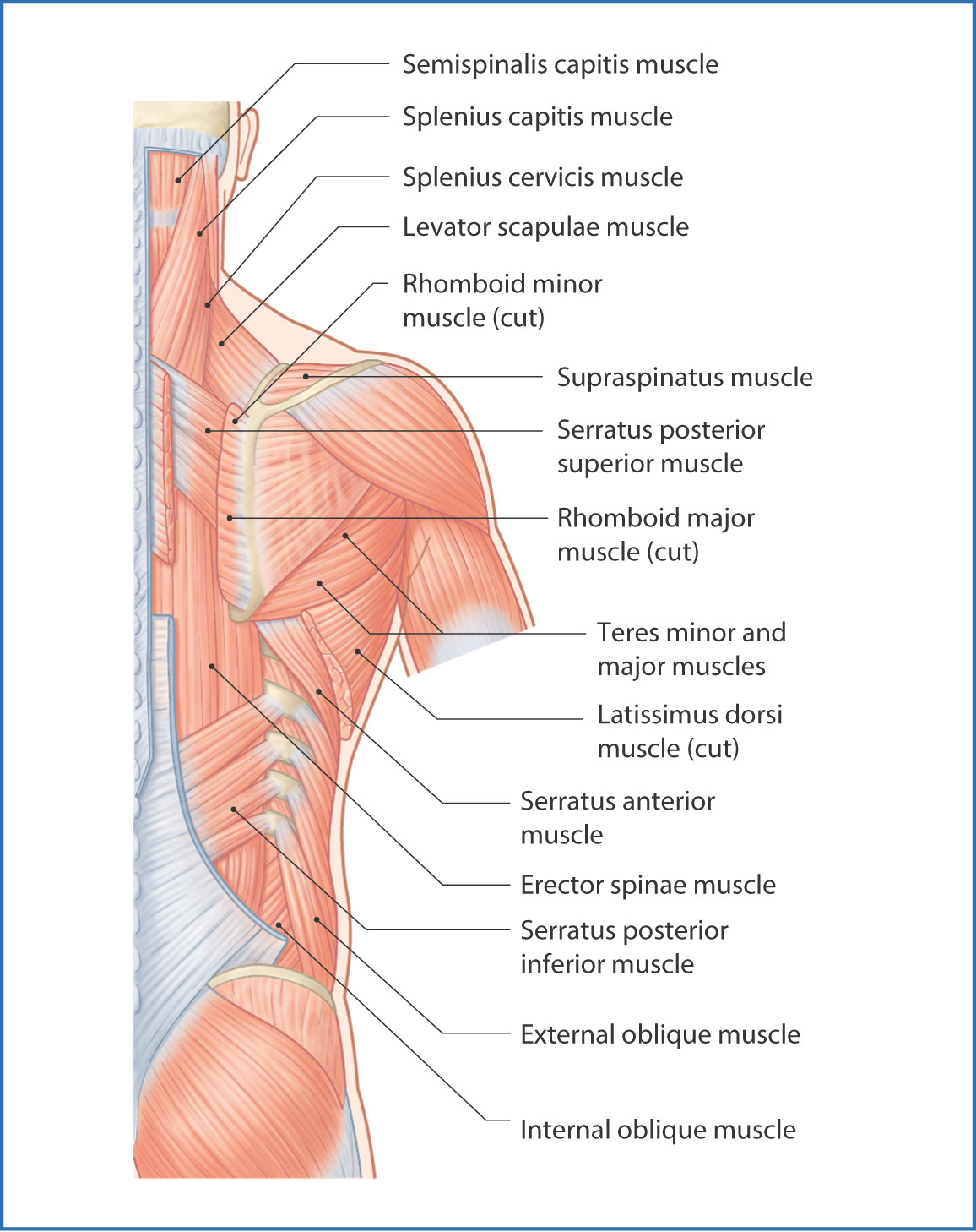
FIGURE 28.2 Intermediate layer of muscles of the back.
- The deep intrinsic back muscles (postvertebral muscles) maintain posture and control movements of the vertebral column and head. These "true" (intrinsic) back muscles are grouped according to their relationship to the surface of the body: the superficial layer of splenius muscles, the intermediate layer of erector spinae muscles (iliocostalis, longissimus, and spinalis), and the deep transversospinales muscles (semispinalis, multifidus, and rotatores) (Fig. 28.3).
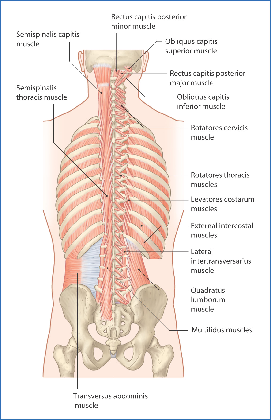
FIGURE 28.3 Deep muscles of the back.
The splenius muscles are divided into capitis and cervicis groups. They originate from the nuchal ligament and spinous processes of TI to TVI and insert onto the superior nuchal line, mastoid process, and posterior tubercles of the transverse processes of CII to CIV. As a group, they extend, laterally flex, and rotate the head and neck.
The intermediate layer of erector spinae muscles forms a broad thick muscle mass that is attached to the sacrum, iliac crest of the pelvis, and lumbar spinous processes. Just inferior to rib XII the group splits into three columns—iliocostalis, longissimus, and spinalis muscles (from lateral to medial). These three columns ascend and insert onto the spinous and transverse processes of more superior vertebrae, ribs, and the posterior base of the skull. They extend the vertebral column and head and assist in rotational and lateral movements of the spine.
The deep or transversospinales muscle group is deep to the erector spinae muscles and runs obliquely from the transverse process of one vertebra to the spinous process of the preceding vertebra. They stabilize, extend, and rotate the vertebral column and include the semispinalis
Jun 11, 2016 | Posted by in ANATOMY | Comments Off on Back Muscles
Order of Human Back Muscles From Superficial to Deep
Source: https://basicmedicalkey.com/back-muscles/
0 Response to "Order of Human Back Muscles From Superficial to Deep"
Publicar un comentario